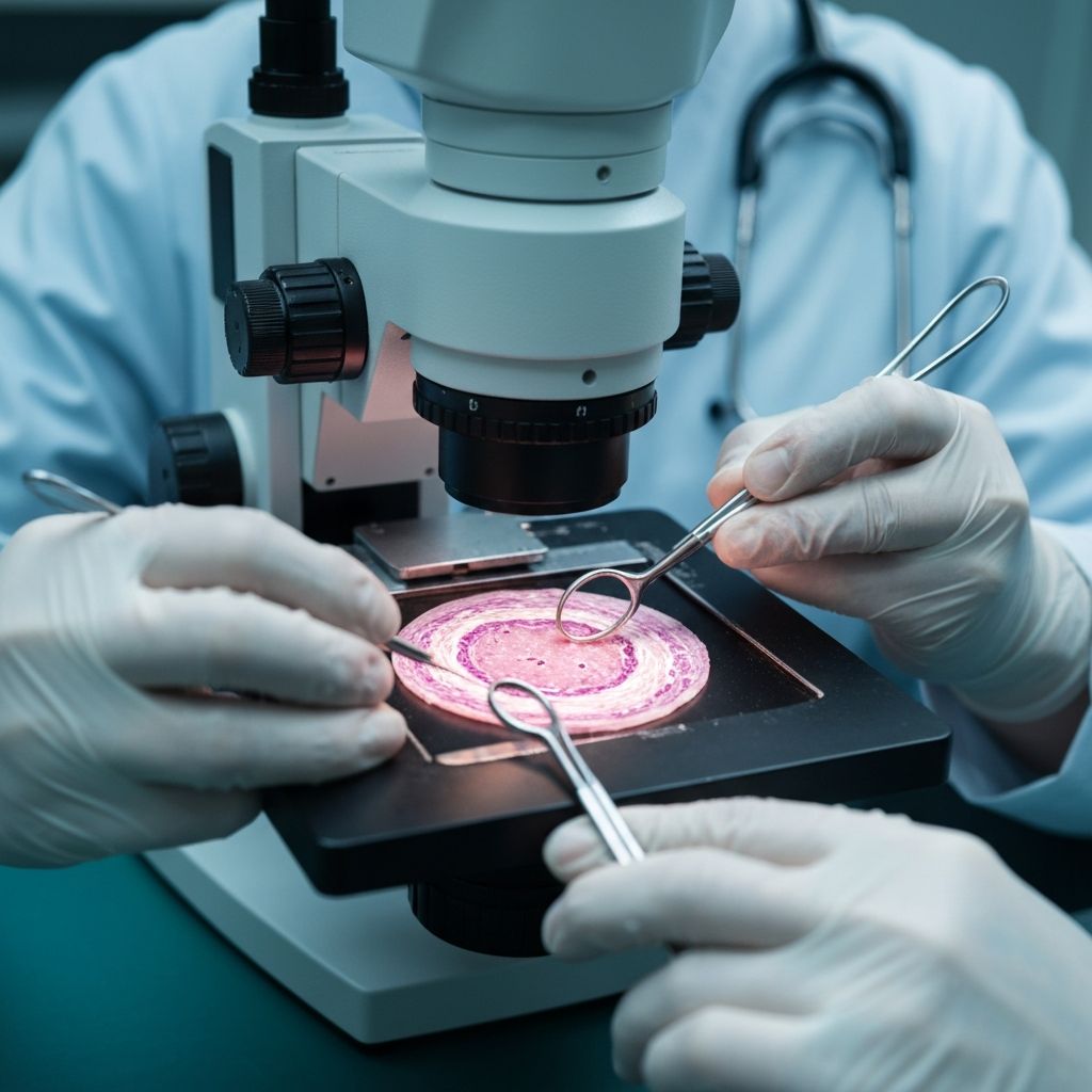
Quantifying a tissue biomarker is not only an image-analysis problem. The reported value can be affected by tissue handling, fixation, staining chemistry, slide preparation, scanning, color management, annotation, thresholding, and the statistical unit of analysis. Reproducibility starts by treating the complete measurement process as the object of evaluation.
Define the measurement before building the pipeline
Specify what is measured, where it is measured, and how the result is reported. A percentage of positive cells, an intensity score, a continuous optical measurement, and a combined score are different quantities. Define the specimen type, stain, tissue compartment, units, cut points, and handling of missing or uninterpretable material before analysis begins.
Control the pre-analytic and staining process
Document fixation time, processing, section thickness, reagent lots, instrument settings, antigen retrieval, controls, and staining run. Include positive and negative controls where appropriate. A stable image pipeline cannot compensate for uncontrolled variation introduced before scanning.
Treat scanning as part of the measurement
Record scanner model, objective, resolution, file format, compression, focus behavior, and color handling. Test representative slides across the scanners and software versions that matter for the intended setting. If a measurement changes after scanning, color normalization, or image conversion, that change belongs in the validation record.
Measure agreement and sources of variation
Use a statistical plan that matches the measurement type. Assess repeatability within a run, reproducibility between runs, readers, scanners, and sites, and agreement near clinically relevant decision thresholds. Report uncertainty and inspect discordant cases rather than relying on a single correlation or agreement statistic.
Biomarker quantification can support research or clinical decision support, but the numeric output is not a diagnosis by itself. Any clinical use requires an appropriate intended use, qualified professional review, and the applicable validation and regulatory pathway.
Sources
- An Image Analysis Solution For Quantification and Determination of Immunohistochemistry Staining Reproducibility (Applied Immunohistochemistry & Molecular Morphology)
- Good Machine Learning Practice for Medical Device Development: Guiding Principles (U.S. Food and Drug Administration)
- Artificial Intelligence Risk Management Framework (National Institute of Standards and Technology)
Written by
Digital Pathology Solutions Editorial Team
Medical AI and digital pathology